Risk of Posttraumatic Stress Disorder and Major Depression in Civilian Patients After Mild Traumatic Brain Injury A TRACK-TBI Study. JAMA Psychiatry 2019

Risk of Posttraumatic Stress Disorder and Major Depression in Civilian Patients After Mild Traumatic Brain Injury A TRACK-TBI Study.

Stein, Sonia Jain, Giacino et al. JAMA Psychiatry. 2019; 76(3):249-258.

https://doi.org/10.1001/jamapsychiatry.2018.4288

Published online: January 30, 2019

Abstract

Importance  Traumatic brain injury (TBI) has been associated with adverse mental health outcomes, such as posttraumatic stress disorder (PTSD) and major depressive disorder (MDD), but little is known about factors that modify risk for these psychiatric sequelae, particularly in the civilian sector.

Objective  To ascertain prevalence of and risk factors for PTSD and MDD among patients evaluated in the emergency department for mild TBI (mTBI).

Design, Setting, and Participants  Prospective longitudinal cohort study (February 2014 to May 2018). Posttraumatic stress disorder and MDD symptoms were assessed using the PTSD Checklist for DSM-5 and the Patient Health Questionnaire-9 Item. Risk factors evaluated included preinjury and injury characteristics. Propensity score weights-adjusted multivariable logistic regression models were performed to assess associations with PTSD and MDD. A total of 1155 patients with mTBI (Glasgow Coma Scale score, 13-15) and 230 patients with nonhead orthopedic trauma injuries 17 years and older seen in 11 US hospitals with level 1 trauma centers were included in this study.

Main Outcomes and Measures  Probable PTSD (PTSD Checklist for DSM-5 score, ≥33) and MDD (Patient Health Questionnaire-9 Item score, ≥15) at 3, 6, and 12 months postinjury.

Results  Participants were 1155 patients (752 men [65.1%]; mean [SD] age, 40.5 [17.2] years) with mTBI and 230 patients (155 men [67.4%]; mean [SD] age, 40.4 [15.6] years) with nonhead orthopedic trauma injuries. Weights-adjusted prevalence of PTSD and/or MDD in the mTBI vs orthopedic trauma comparison groups at 3 months was 20.0% (SE, 1.4%) vs 8.7% (SE, 2.2%) (P < .001) and at 6 months was 21.2% (SE, 1.5%) vs 12.1% (SE, 3.2%) (P = .03). Risk factors for probable PTSD at 6 months after mTBI included less education (adjusted odds ratio, 0.89; 95% CI, 0.82-0.97 per year), being black (adjusted odds ratio, 5.11; 95% CI, 2.89-9.05), self-reported psychiatric history (adjusted odds ratio, 3.57; 95% CI, 2.09-6.09), and injury resulting from assault or other violence (adjusted odds ratio, 3.43; 95% CI, 1.56-7.54). Risk factors for probable MDD after mTBI were similar with the exception that cause of injury was not associated with increased risk.

Conclusions and Relevance  After mTBI, some individuals, on the basis of education, race/ethnicity, history of mental health problems, and cause of injury were at substantially increased risk of PTSD and/or MDD. These findings should influence recognition of at-risk individuals and inform efforts at surveillance, follow-up, and intervention.

Sport-related Concussion Clinical Profiles: Clinical Characteristics, Targeted Treatments, and Preliminary Evidence. Curr Sports Med Rep 2019

Sport-related Concussion Clinical Profiles: Clinical Characteristics, Targeted Treatments, and Preliminary Evidence.

Kontos, Sufrinko, Sandel, Emami, Collins. Curr Sports Med Rep. 2019 Mar;18(3):82-92.

www.doi.org/10.1249/JSR.0000000000000573

Abstract

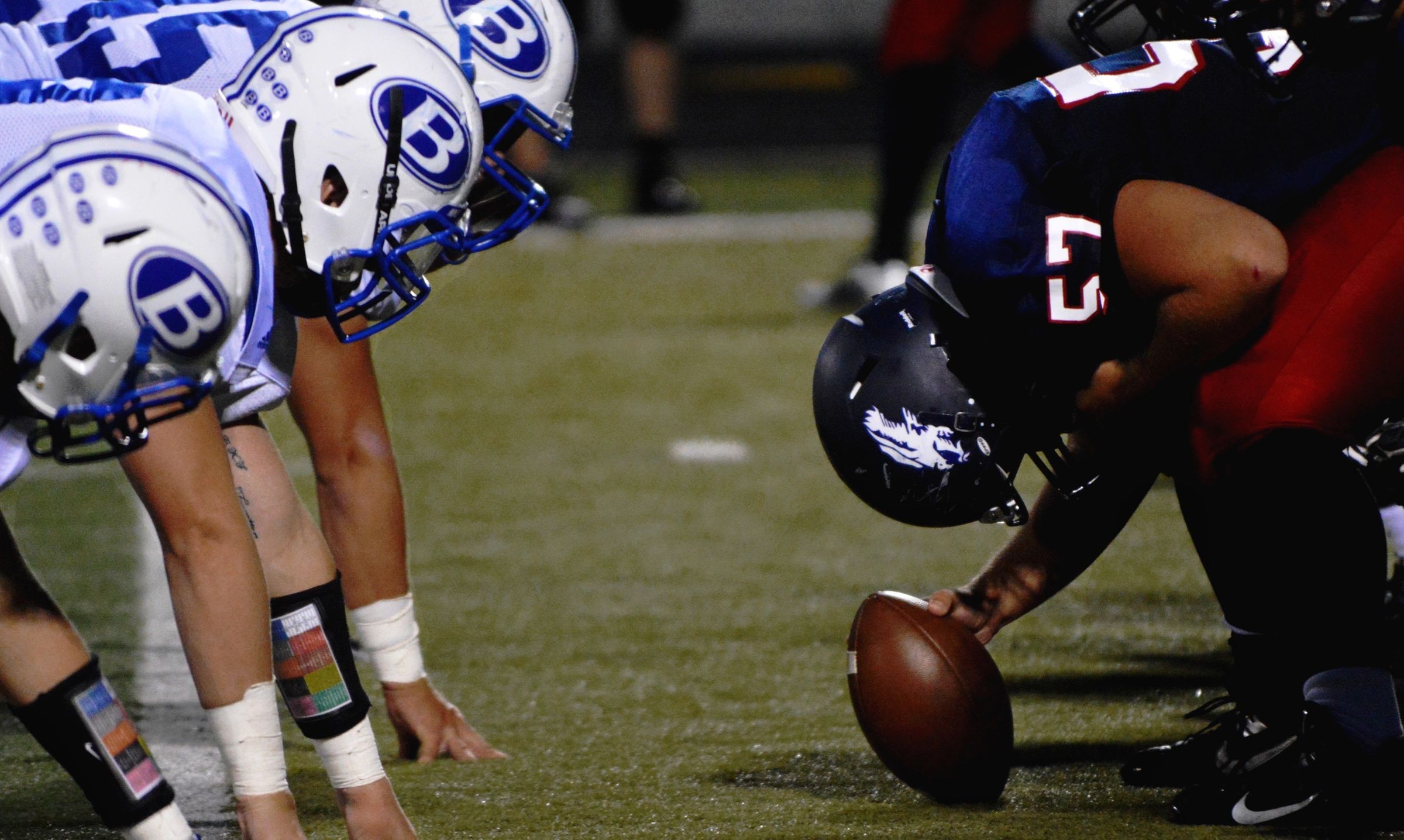
Sport-related concussion (SRC) is a heterogeneous injury that involves varied symptoms and impairment that presents a significant clinical challenge to sports medicine professionals.

In response to this challenge, clinical researchers have proposed clinical profiles or subtype models for assessing and treating athletes with SRC. One such model emphasizes five concussion clinical profiles including cognitive/fatigue, vestibular, ocular, migraine, and anxiety/mood. Sleep is a common modifier that co-occurs across these clinical profiles. A combination of medical history, risk factors, injury information, clinical characteristics, and assessment outcomes can inform each clinical profile.

Preliminary data involving 236 patients from a concussion specialty clinic indicate that the migraine (26%) and anxiety/mood (24%) profiles are the most common, with vestibular and ocular profiles combined representing more than one third (35%) of clinical profiles.

Findings also support several relationships among different clinical profiles including vestibular and migraine, suggesting that many athletes present with multiple clinical profiles. Targeted, active treatments for each profile are discussed.

Combining H-FABP and GFAP increases the capacity to differentiate between CT-positive and CT-negative patients with mild traumatic brain injury. PLoS ONE, 2019

Lagerstedt L, Egea-Guerrero JJ, Bustamante A, Rodríguez-Rodríguez A, El Rahal A, Quintana-Diaz M, et al. (2018) Combining H-FABP and GFAP increases the capacity to differentiate between CT-positive and CT-negative patients with mild traumatic brain injury. PLoS ONE 13(7): e0200394.

https://doi.org/10.1371/journal.pone.0200394

Abstract:

Mild traumatic brain injury (mTBI) patients may have trauma-induced brain lesions detectable using CT scans. However, most patients will be CT-negative. There is thus a need for an additional tool to detect patients at risk. Single blood biomarkers, such as S100B and GFAP, have been widely studied in mTBI patients, but to date, none seems to perform well enough. In many different diseases, combining several biomarkers into panels has become increasingly interesting for diagnoses and to enhance classification performance.

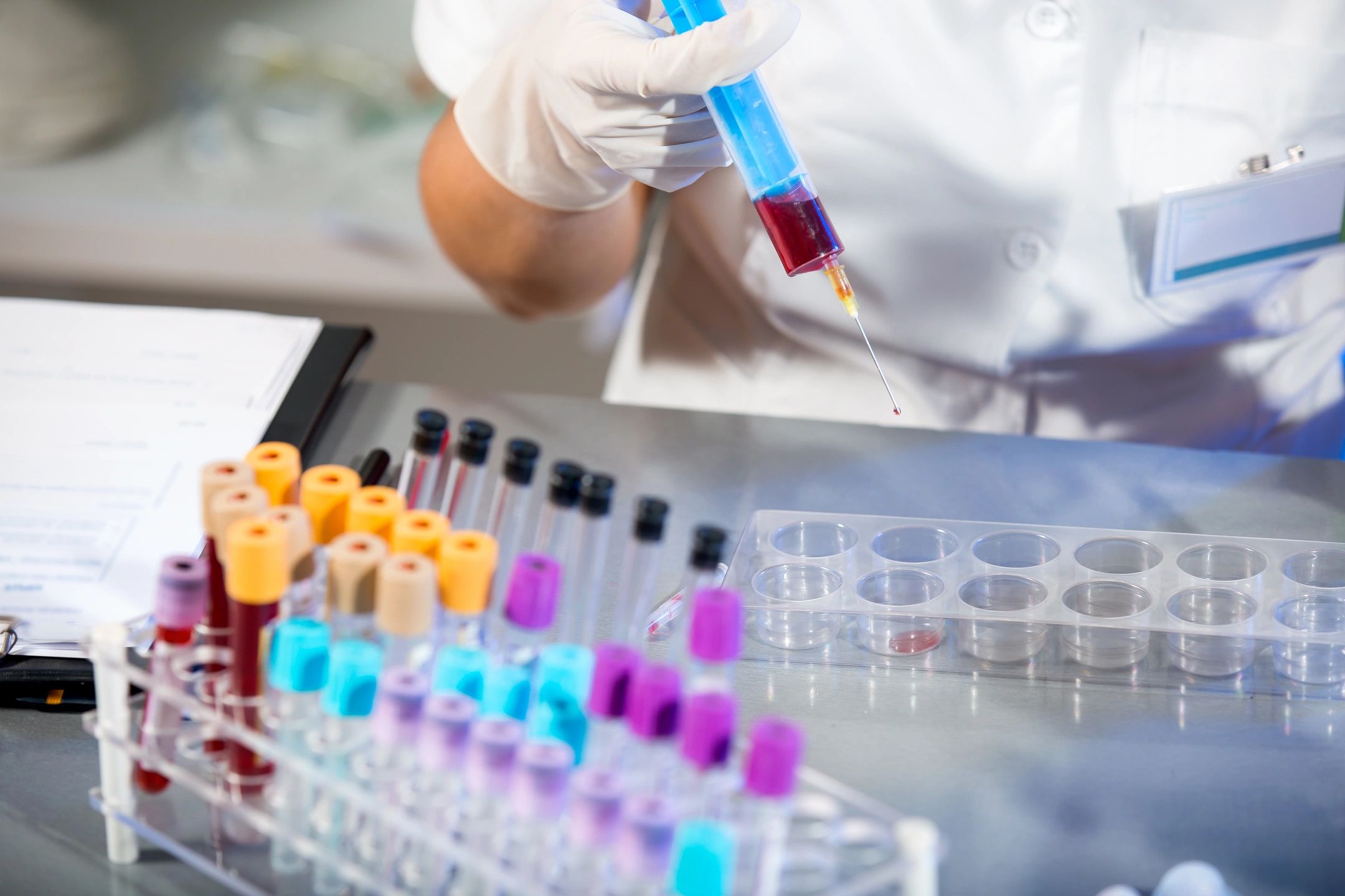
The present study evaluated 13 proteins individually—H-FABP, MMP-1, MMP-3, MMP-9, VCAM, ICAM, SAA, CRP, GSTP, NKDA, PRDX1, DJ-1 and IL-10—for their capacity to differentiate between patients with and without a brain lesion according to CT results. The best performing proteins were then compared and combined with the S100B and GFAP proteins into a CT-scan triage panel. Patients diagnosed with mTBI, with a Glasgow Coma Scale score of 15 and one additional clinical symptom were enrolled at three different European sites. A blood sample was collected at hospital admission, and a CT scan was performed. Patients were divided into two two-centre cohorts and further dichotomised into CT-positive and CT-negative groups for statistical analysis. Single markers and panels were evaluated using Cohort 1.

Four proteins—H-FABP, IL-10, S100B and GFAP—showed significantly higher levels in CT-positive patients. The best-performing biomarker was H-FABP, with a specificity of 32% (95% CI 23–40) and sensitivity reaching 100%. The best-performing two-marker panel for Cohort 1, subsequently validated in Cohort 2, was a combination of H-FABP and GFAP, enhancing specificity to 46% (95% CI 36–55). When adding IL-10 to this panel, specificity reached 52% (95% CI 43–61) with 100% sensitivity.

These results showed that proteins combined into panels could be used to efficiently classify CT-positive and CT-negative mTBI patients.

Facebook
Twitter
LinkedIn